Nail Intramedullari Best Practices and Techniques?
nail intramedullari techniques have gained significant attention in orthopedic practices. They serve as a vital tool for treating bone fractures. The effectiveness of these methods often relies on precise application.
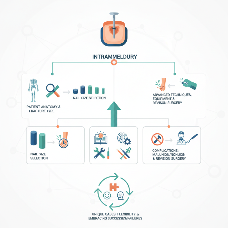
Surgeons must consider various factors, including patient anatomy and the type of fracture. Choosing the right nail size is crucial. This decision impacts healing and recovery times. Misjudging this can lead to complications like malunion or nonunion. These issues require skilled revision surgery and can prolong patient suffering.
Despite advancements, challenges remain. Surgeons often express a need for improved techniques and equipment. Continuous learning is essential. There is no one-size-fits-all approach to nail intramedullari procedures. Each case is unique, demanding flexibility and judgment. Embracing both successes and failures enriches the practice and enhances overall outcomes.
Overview of Nail Intramedullari Fixation Techniques
Intramedullary nailing has become a popular fixation technique for bone fractures. This method involves inserting a rod inside the medullary cavity of the bone. It stabilizes the fracture, allowing for quicker recovery. Surgeons often prefer this technique for its minimally invasive approach. However, mastering the procedure requires skill and practice.
Surgeons must consider various factors when choosing the right technique. For instance, the type of fracture influences the choice of nail and insertion technique. Additionally, patient-specific factors like age and bone quality play a crucial role. Anatomical variations can complicate insertion. Thus, a deep understanding of the anatomy is essential for success.
One challenge in intramedullary nailing is achieving the correct alignment. Misalignment can lead to complications. Surgeons must remain vigilant during the procedure. Even experienced surgeons encounter unexpected issues. Continuous reflection and adaptation are necessary to improve outcomes. In conclusion, the journey of mastering intramedullary fixation involves balancing technique precision with the unpredictability of real-world cases.
Indications for Nail Intramedullari Procedures
Intramedullary nailing is a common technique in orthopedic surgery for treating bone fractures, particularly in the long bones of the legs. This method is preferred for its ability to stabilize fractures while preserving the fracture site's vascular supply. Indications for intramedullary nailing procedures mainly include diaphyseal fractures of the femur and tibia. These fractures often result from trauma, and timely intervention is crucial for optimal recovery.
In addition to these fractures, intramedullary nailing is indicated for certain pathological conditions. For instance, metastatic bone disease or benign tumors may require stabilization. In these cases, the choice of nail type and insertion technique becomes critical. The surgeon must consider factors like the patient's age, activity level, and overall health as they choose the appropriate approach.
While effective, there are challenges that come with intramedullary nailing. Complications such as non-unions and malalignments may occur. Surgeons need to reflect on these issues and adjust techniques accordingly. Surgical precision is essential, and sometimes, complications arise due to anatomical variations or unexpected patient responses. This reinforces the need for careful preoperative planning and precise execution during the procedure.
Step-by-Step Surgical Procedure for Nail Intramedullari Placement
The placement of intramedullary nails is a critical procedure in orthopedic surgery. This technique stabilizes fractures in long bones. The key to success lies in meticulous preparation and execution. Surgeons must assess the fracture type accurately. Planning is essential for optimal nail selection and positioning.
During the procedure, anesthesia is administered to ensure patient comfort. The surgical site is then marked and cleaned. A small incision is made above the fracture. A reamer is used to create a pathway for the nail. It's important to maintain a precise angle during insertion. Surgeons often face challenges, such as aligning the nail correctly. A misstep can lead to complications, urging the need for careful consideration at every stage.
Once the nail is in place, fixation is crucial. The use of locking screws provides added stability. However, achieving perfect alignment may be difficult. Post-operative imaging checks are vital to assess the outcome. Reflection on technique can help improve future procedures. Each case offers valuable lessons and opportunities for growth in surgical practice.
Postoperative Care and Rehabilitation Strategies
Postoperative care after nail intramedullary fixation is crucial for optimal recovery. A study highlights that adherence to rehabilitation protocols significantly reduces complications. Approximately 20% of patients experience delayed healing if not managed properly. Early mobilization is essential, yet patients often hesitate to begin rehabilitation. This reluctance can lead to stiffness and limited range of motion, making recovery slower.
Physical therapy plays a vital role in enhancing recovery outcomes. Patients are encouraged to engage in tailored exercises within two weeks post-surgery. The goal is to regain strength and improve functionality. A report indicates that structured rehabilitation can increase return to daily activities by 30%. However, many patients may not fully commit to their rehabilitation plan. They might overlook simple exercises due to pain or fear of reinjury. This can hinder full recovery, highlighting the need for better patient education.
Monitoring progress is equally important. Regular follow-up sessions help to address concerns and adjust rehabilitation strategies. Studies reveal that patients who attend follow-ups have a 40% better recovery rate. However, some individuals may not prioritize these visits, risking their rehabilitation success. Open communication between patients and healthcare providers can enhance motivation and ensure proper recovery.
Common Complications and Management Approaches in Nail Intramedullari Surgery
Intramedullary nailing is a common procedure for long bone fractures. While effective, it can lead to complications. Infection, non-union, and malalignment are prominent issues. A study showed that up to 20% of patients face complications post-surgery. These risks can be attributed to various factors, including surgical technique and patient health.
Effective management is crucial. Infection is a significant concern. Prophylactic antibiotics may reduce risk. Regular follow-ups are essential to monitor healing. Non-union can present challenges too. This requires careful assessment and, sometimes, additional procedures. An early diagnosis can significantly improve outcomes.
Tip: Proper alignment during surgery is vital. Use imaging to guide placement. Monitor the patient’s overall health. This can make a difference in recovery. Complications are a reality. Reflecting on past cases can lead to better techniques. Every injury is unique; adjust your approach accordingly.